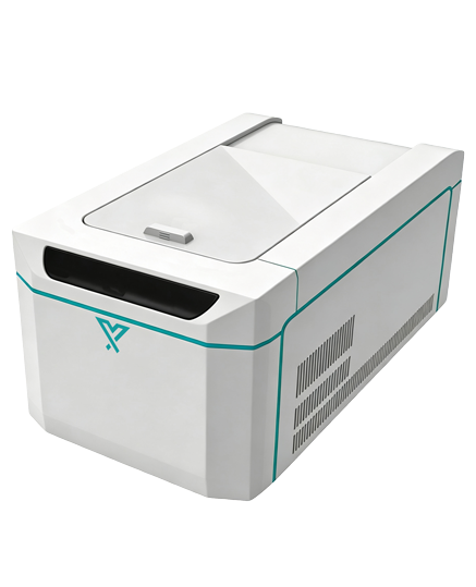
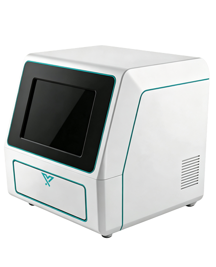
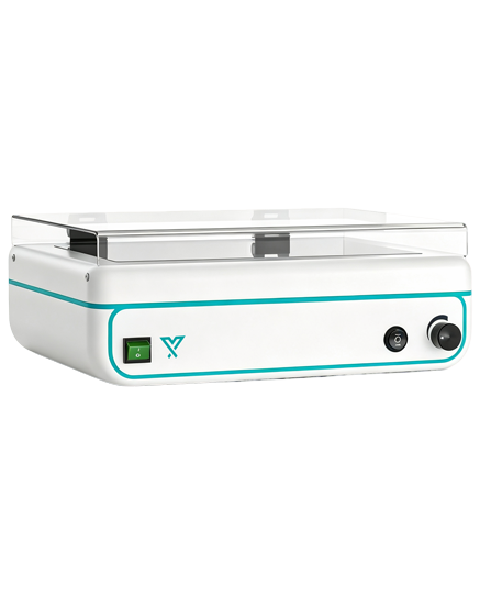
1. Automated Imaging Workflow
Pre‑configured imaging parameters are available for nucleic acid gels (EB, SYBR Green, SYBR Safe) and protein gels (Coomassie Brilliant Blue, silver staining). Users only need to load samples; the software automatically executes focusing, exposure, image optimization and band detection. Standardized “load‑image‑export” operation minimizes human‑induced errors.
2. Research‑Grade CCD Camera
Built‑in high‑sensitivity 6‑megapixel CCD detector features low dark current and 16‑bit analog‑to‑digital conversion. Coupled with a high‑transmittance fixed‑focus lens, it clearly captures weak‑positive bands and low‑abundance samples. Its broad dynamic range ensures both strong and faint signals are well‑represented in a single image.
3. Multi‑Wavelength UV Excitation Light Source
The standard 302 nm mid‑wave UV transilluminator is compatible with common nucleic acid dyes such as EB and SYBR Green. Stepless adjustable light intensity adapts to gels with different staining levels to prevent over‑exposure or under‑exposure.
4. Fully‑Enclosed Dark Chamber with Pull‑Out Sample Stage
The dark chamber effectively blocks ambient stray light for clean imaging background. The smoothly pull‑out stage accommodates large‑size gels (up to 20 × 20 cm) or membranes, and supports multiple sample holders.
5. Integrated Image Analysis Software
Dual‑module software for image acquisition and analysis is included. The acquisition module provides auto‑exposure, auto‑white balance and real‑time preview. The analysis module supports automatic band recognition, molecular‑weight ladder fitting, grayscale quantification, colony counting and more. Data can be exported to Excel, PDF or report templates to meet routine laboratory data‑management requirements.