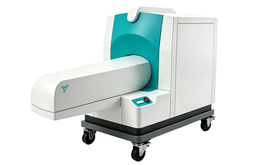
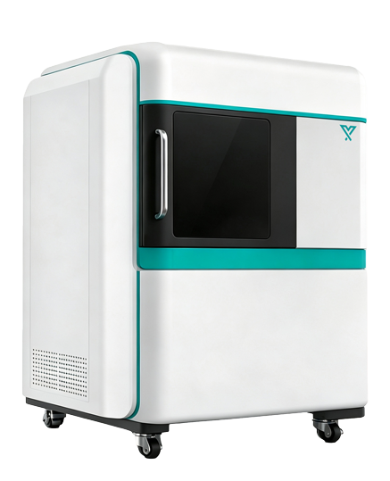
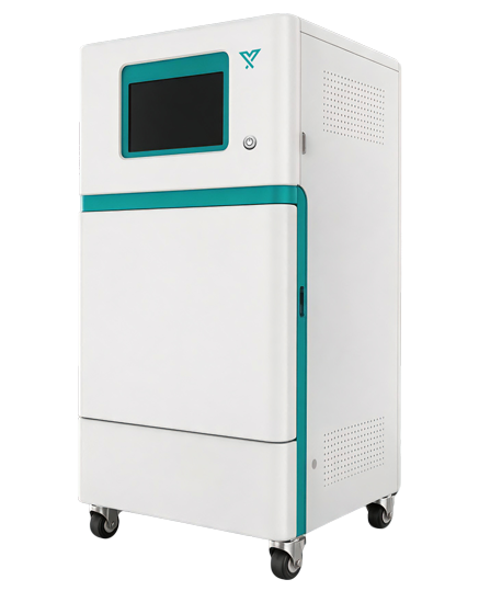
1. High‑Sensitivity 2D Optical Imaging
An approximately 1‑megapixel scientific‑grade cooled camera is paired with a large‑aperture motorized lens to capture BLI or FLI signals and perform 2D quantification. Equipped with 20 monochromatic LED light sources across 24 excitation positions, as well as up to 18 narrow‑band emission filters of various wavelengths covering visible‑light to NIR‑I, it enables fluorescence signal detection for diverse fluorescent proteins and fluorescent probe labels. Light intensity and imaging channels can be automatically invoked by software to minimize manual adjustments. The 24‑position motorized filter wheel automatically switches emission channels according to experimental protocols.
2. Micro‑Focus X‑ray Source
The maximum X‑ray tube voltage reaches approximately 90 kV. Tube voltage, tube current and exposure time can be adjusted according to animal species, tissue density and scanning objectives. The micro‑focus design preserves structural details under high‑magnification scanning.
3. High‑Sensitivity Flat‑Panel Detector
The high‑resolution flat‑panel detector adopts low‑noise readout and high‑dynamic‑range imaging to acquire X‑ray projection data from multiple angles, laying the foundation for 3D tomographic reconstruction.
4. 360° Rotation Stage
A high‑precision rotation stage drives animals or samples for multi‑angle projection acquisition. Multiple fields‑of‑view and magnification levels allow trade‑offs among scanning coverage, spatial resolution and scan duration.
5. Multiple Scanning Modes
Fast preview, low‑dose in‑vivo scanning and high‑resolution scanning are supported. Users may select corresponding scanning protocols for longitudinal animal studies, ex‑vivo bones or material samples.
6. Animal Anesthesia and Temperature Control
The stage integrates anesthesia input, waste‑gas recovery and animal temperature‑keeping functions. Interfaces for respiratory or physiological monitoring are reserved to minimize animal motion and body‑temperature drop during long‑duration scans.
7. Low‑Dose for Longitudinal Observation
Optimized exposure parameters, projection counts and reconstruction settings reduce unnecessary radiation dose, enabling repeated scans of the same animal at multiple time points throughout the study period.
8. Radiation Protection and Safety Interlock
Shielding enclosure, chamber‑door interlock, status indicators and emergency‑stop devices jointly ensure operational safety. The system monitors the real‑time operating status of the X‑ray source and key components.
9. 3D Reconstruction and Quantitative Analysis
The software supports tomographic reconstruction, 2D slice viewing, MPR multi‑planar reconstruction, 3D volume rendering and region segmentation. Further quantitative analysis is available for bone mineral density, bone volume fraction, trabecular thickness, trabecular number, trabecular separation and tissue volume.